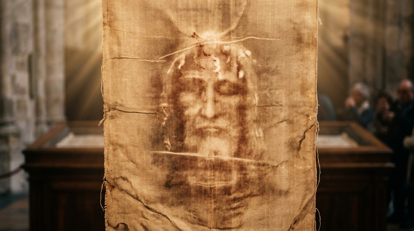
Forensic Autopsy

Forensic Pathology & Blood Chemistry

Every mark on the Shroud corresponds with precision to Roman crucifixion practices, forensic traumatology, and genuine physiological reactions that no artist before the 20th century could have conceived.

STURP Hematology

Real Human Blood (Type AB)

Heller & Adler's Definitive Chemical Proof

In 1980, STURP biophysicists Dr. John Heller (New England Institute) and Dr. Alan Adler (Western Connecticut State University), recognized world authorities on porphyrin chemistry, subjected the blood marks to comprehensive chemical testing:

  • Positive for Hemoglobin: Generated characteristic hemochromogen crystals, positive for porphyrin and protein tests.
  • Primate Albumin & Antibodies: Immunological tests confirmed human primate origin.
  • High Bilirubin Levels: Explains why the blood stains remain reddish rather than turning dark brown; high bilirubin is directly produced by severe traumatic stress and crushing hemolysis.
  • Blood Transferred First: Proteolytic enzyme tests proved the blood was clotted on the cloth before the image formed. Beneath the blood clots, the linen fibers are entirely unoxidized.
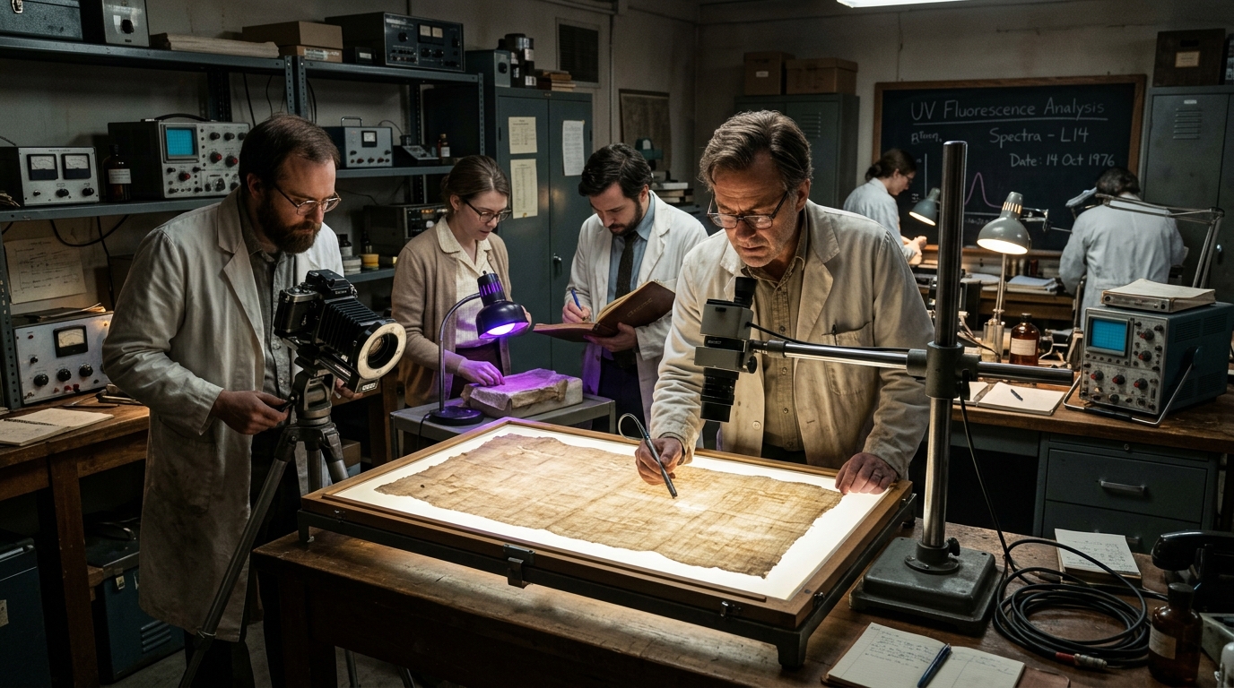
Spectroscopic Blood Analysis of Shroud of Turin
Type AB Human Blood
Trauma Evidence

Six Incontrovertible Forensic Facts

Forensic pathologists, including Dr. Pierre Barbet, Dr. Robert Bucklin (Los Angeles County Deputy Medical Examiner), and Dr. Anthony Sava, have documented:

1. Destot's Space

Nails in the Wrists, Not Palms

All medieval art depicted nails through the open palms. Forensic experiments proved that palms tear through under body weight. The Shroud depicts the nail driven through Destot's space in the wrist, causing the median nerve to retract the thumbs inward.

2. Roman Scourging

Flagrum Taxillatum Contusions

Over 120 dumbbell-shaped contusions cover the shoulders, back, buttocks, and calves. Their angles demonstrate two lictors: one on the left, one on the right, with differing statures executing the brutal Roman flagellation.

3. Helmet of Thorns

Arterial & Venous Scalp Bleeding

Punctures cover the entire scalp (not just a decorative circlet). The blood trickles along the forehead furrows with distinct pulsatile arterial flows and slower venous drips, adhering to gravity as the victim moved his head.

4. Post-Mortem Rigor Mortis

Crucifixion Asphyxiation Stance

The body exhibits severe post-mortem rigidity (rigor mortis). The ribcage is expanded in an extreme inspiratory state, characteristic of death by suspension asphyxiation where the victim struggled upward to exhale.

5. The Lance Wound

Blood and Water Separation

An elliptical wound between the 5th and 6th ribs released settled red blood cells followed by clear pericardial and pleural serous fluid (John 19:34). UV fluorescence reveals a serum halo invisible to the naked eye.

6. No Decomposition

Body Removed Within 36–40 Hours

There are zero signs of putrefaction or bacterial decay on the linen. Yet, rigor mortis is established and blood clotted. Pathologists conclude the body remained in the cloth for a minimum of 24 hours and a maximum of 40 hours.

Interactive Tool

Click Wound Points for Forensic Clinical Details

Select any pin on the anatomical figure below to review the medical and scriptural observations.

Forensic Hotspots of the Shroud
Forensic Wound Analysis

Facial Trauma & Contusions

Clinical Observation

Swollen right cheek, deviation of the nasal cartilage consistent with a violent blow from a club or rod, lacerations along the eyebrow and forehead, swollen lip, swollen eye.

Scriptural Correlation

John 19:3 - "And they struck him with their hands."

Forensic Significance

Maxillofacial surgeons note realistic soft tissue swelling and hematoma formation that defies artistic stylization.