Unique 3D VP-8 Topography
Cloth-to-Body Distance Encoded in Luminance
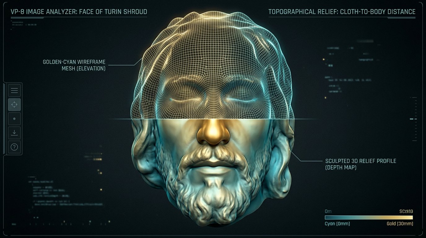
In 1976, NASA scientists John Jackson and Eric Jumper discovered that the Shroud image contains true three-dimensional topographic information. Normal photographs or paintings flatten or distort when placed in a VP-8 Image Analyzer; the Shroud creates a anatomically perfect 3D relief.
Key Scientific Takeaways:
- Luminance values correspond directly to the distance between the cloth and the body.
- No artist in history has ever painted or photographed with mathematical distance encoding.
- Reflectance gradients indicate an orthogonal (straight-line) collimated emission of energy.
- Yields anatomically accurate facial contours, nose bridge, eye sockets, and facial bone structure.
Microscopic & Chemical Impossibility
Oxidation of the 200nm Primary Cell Wall Only
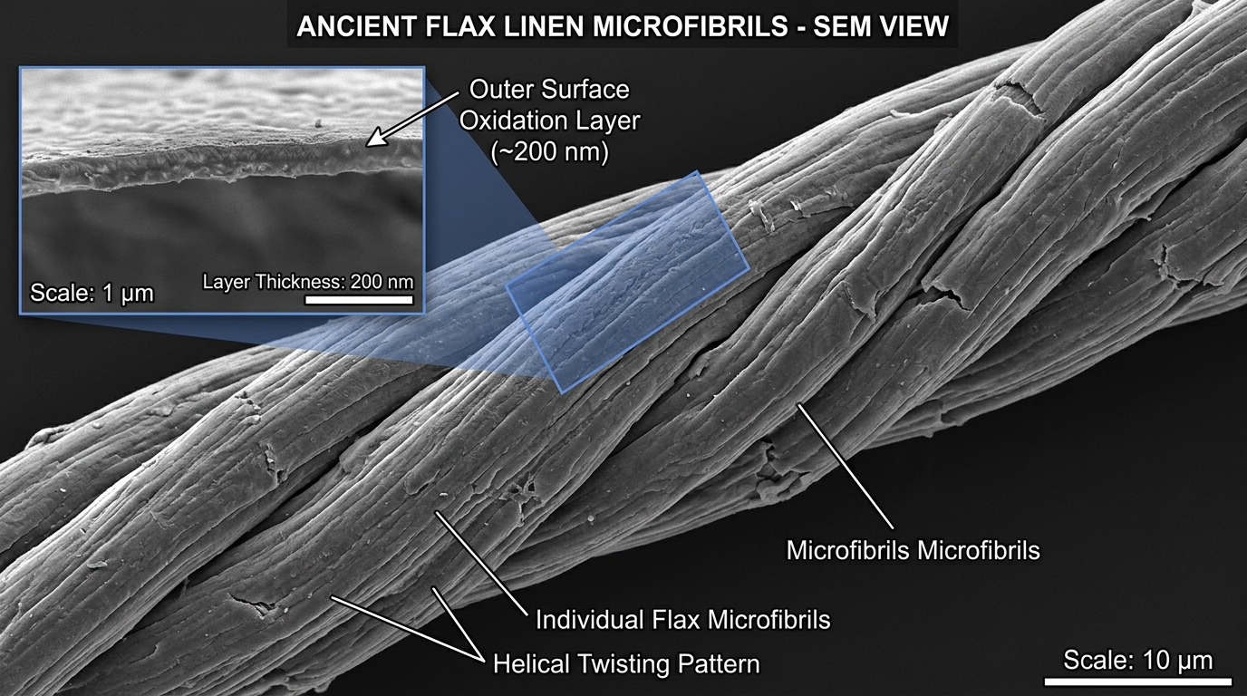
The 1978 STURP (Shroud of Turin Research Project) spent over 120 continuous hours running non-destructive physical and chemical tests. They proved definitively: there is zero paint, dye, pigment, brushstrokes, or binder. The image is an ultra-thin discoloration confined strictly to the topmost 200–500 nanometers of the linen microfibrils.
Key Scientific Takeaways:
- The discoloration is caused by the oxidation and dehydration of the linen's primary cellulose wall.
- The coloration does not penetrate the medulla or inside of the linen fiber—it is razor-thin (1/100th of a human hair).
- No capillary action or capillary flow of liquids occurred (conclusively ruling out liquids, oils, or paints).
- Italian ENEA researchers (2011) discovered the only known physical mechanism to duplicate this depth and coloration is short-duration, high-intensity vacuum ultraviolet (VUV) radiation.
Forensic Pathology & Real Human Blood
Type AB Blood, Pre-Image Contact, & Flawless Anatomy
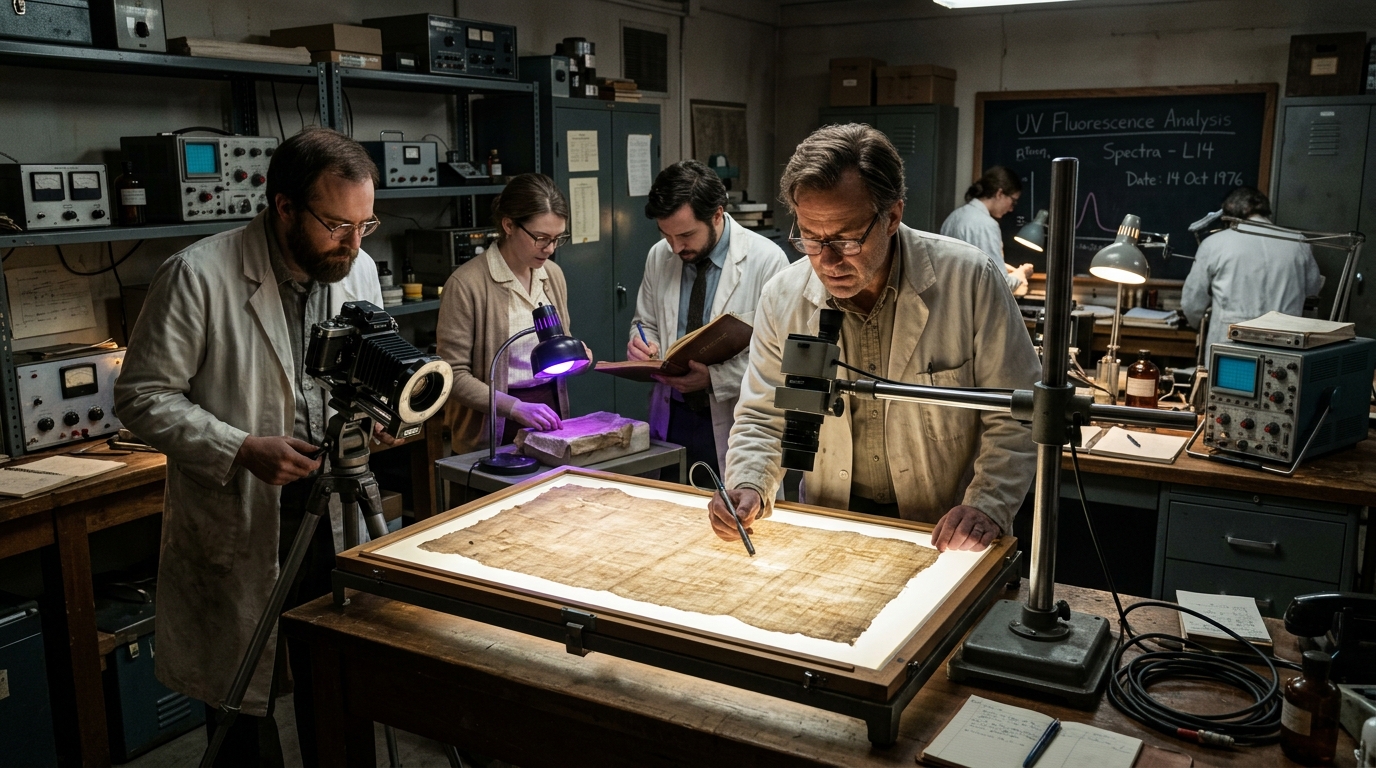
Biophysicists John Heller and Alan Adler proved the stains are real human blood—positive for hemoglobin, human albumin, bilirubin, and high serum. Strikingly, tests confirm the blood went onto the cloth FIRST; the image was subsequently formed around and between the clotted blood without affecting the cloth beneath.
Key Scientific Takeaways:
- Blood Type AB identified with positive human primate antigens and XY male chromosomal components.
- Anatomical accuracy unknown in medieval Europe: nails through Destot's space in the wrist (not the palms), causing reflex retraction of the thumbs.
- Over 120 dumbbell-shaped scourge marks matching the Roman flagrum taxillatum, delivered from two whip-bearers of different heights.
- Halo of serum albumin visible only under UV fluorescence around the post-mortem side wound and blood clots.
- Post-mortem rigor mortis and chest expansion typical of death by crucifixion and asphyxiation.
The 1988 C-14 Debunked & 2022 WAXS Dating
Invisible Reweave Discovered & X-Ray Crystallography Matches 1st Century
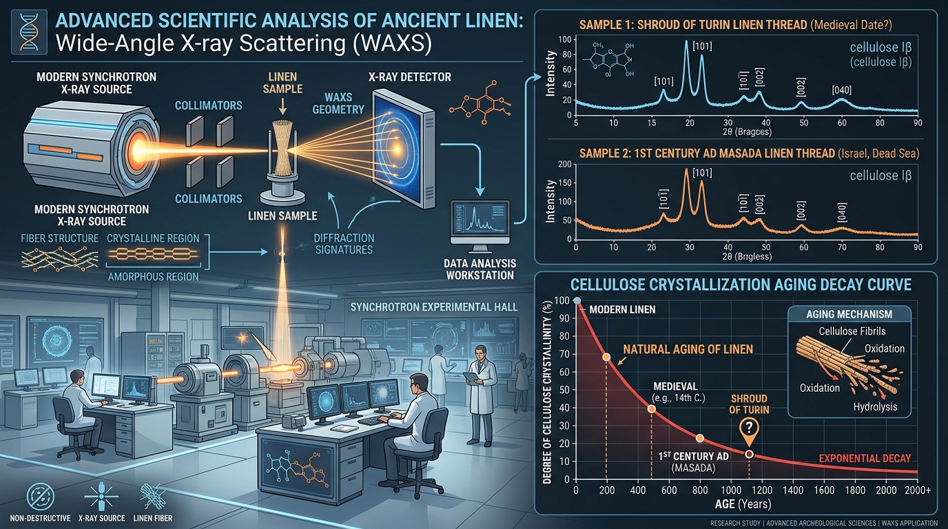
The infamous 1988 Carbon-14 test dating the Shroud to 1260–1390 was completely compromised. Los Alamos chemist Raymond Rogers proved in 2005 that the test sample came from a 16th-century invisible repair containing cotton and madder dye. In 2022, Italian crystallographer Dr. Liberato De Caro utilized Wide-Angle X-ray Scattering (WAXS) to date the linen cellulose directly to the 1st century AD.
Key Scientific Takeaways:
- 1988 corner sample contained anomalous cotton fibers, gum-dye mordant, and low vanillin, absent in the rest of the Shroud.
- Raymond Rogers published in peer-reviewed Thermochimica Acta (2005) proving the sample was chemically unrepresentative.
- 2019 legal release of raw 1988 laboratory data revealed statistical heterogeneity, proving the C-14 result was invalid according to international metrology standards.
- Dr. Liberato De Caro (CNR Institute of Crystallography, 2022) measured cellulose structural aging via WAXS: dated directly to ~2000 years, exactly matching 1st-century AD linen from the siege of Masada.
Botanical & Mineral Fingerprints
Jerusalem Pollen & Rare Travertine Aragonite Limestone
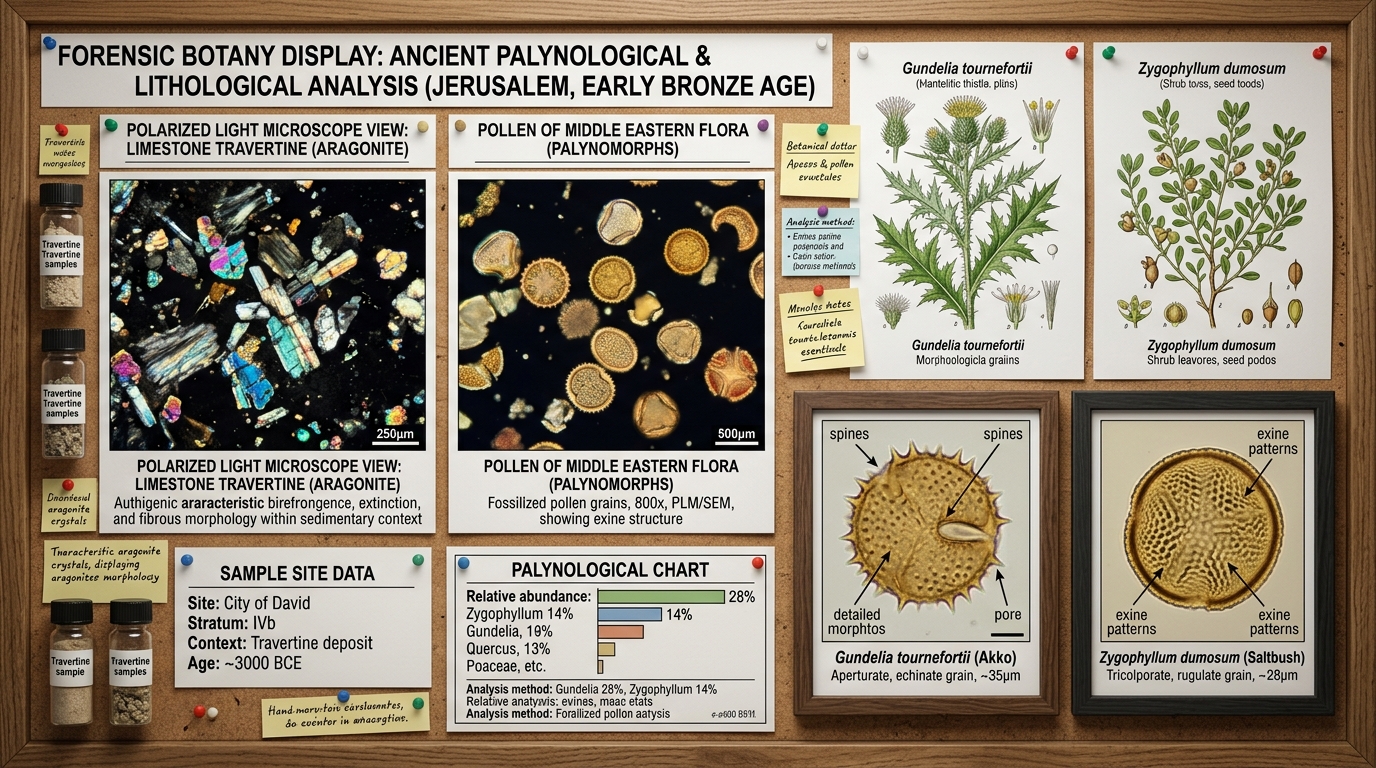
Swiss criminologist Dr. Max Frei and Hebrew University botanist Prof. Avinoam Danin identified over 58 distinct pollen types on the Shroud. A heavy concentration belongs to plants endemic only to the Judean desert and Jerusalem area during spring. Furthermore, microscopic limestone particles found on the feet, knees, and nose match the rare travertine aragonite of ancient Jerusalem tombs.
Key Scientific Takeaways:
- Pollen grains from Gundelia tournefortii (a desert thorn) and Zygophyllum dumosum, plants that co-exist exclusively in the Jerusalem-Dead Sea corridor.
- High concentration of Gundelia pollen suggests a fresh plant was placed near the head in spring (Passover timeframe).
- Spectrographic analysis of dirt on the feet and nose matches travertine aragonite limestone from ancient quarries outside Damascus Gate in Jerusalem.
- Invisible to the naked eye; impossible for a medieval European forger to conceive, collect, or forge.
The Sister Cloth: Sudarium of Oviedo
7th-Century Documented Face Veil with Over 90% Blood Geometry Congruence
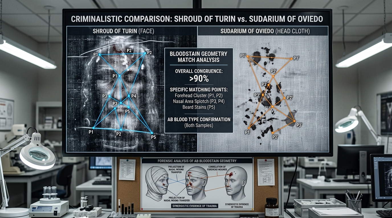
The Sudarium of Oviedo is an ancient face cloth kept in northern Spain with an unbroken documented provenance dating back to 614 AD—over 600 years BEFORE the 1988 C-14 medieval date. Criminalistic analysis demonstrates that both cloths wrapped the exact same human head immediately following death.
Key Scientific Takeaways:
- Documented in Oviedo, Spain since the 7th century, safely stored in the Arca Santa.
- Identical rare AB blood group, identical nasal pulmonary edema fluid ratio from suffocation.
- Geometric polar coordinate overlays by the Spanish Centre for Sindonology (EDICES) reveal over 70 matching facial wound points (>90% correlation).
- Pollen types on the Sudarium mirror the Shroud's Jerusalem flora (Gundelia tournefortii).
- Because the Sudarium has an ironclad pre-7th century history, its forensic match to the Shroud physically disproves any medieval origin.